



Articolo del 29/07/2026
Undergoing a Magnetic Resonance Imaging (MRI) scan can feel intimidating, yet it remains one of the most extraordinary and safe modalities in modern medicine. Unlike X-rays or CT scans, an MRI involves zero ionizing radiation. Instead, it harnesses powerful magnetic fields and radio waves to generate remarkably detailed images of soft tissues, organs, and bones.
To visualize how it works, imagine the human body as a vast collection of tiny magnets (hydrogen atoms). The MRI scanner temporarily aligns these atoms and gently “questions” them with radio pulses. As the atoms respond, state-of-the-art computers translate their signals into high-resolution, three-dimensional diagnostic maps.
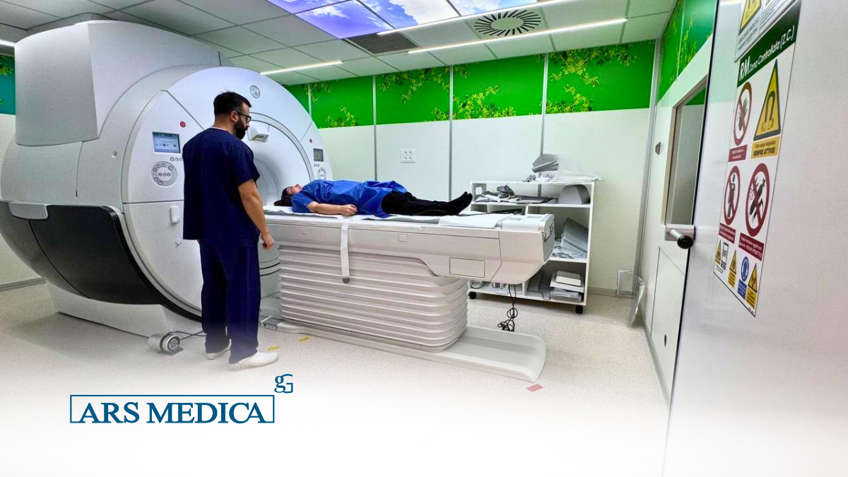
While traditional MRI is already an outstanding diagnostic tool, the 3 Tesla (3T) system—available here at Ars Medica—represents an essential leap forward when millimeter-level precision can alter the course of treatment. It is not simply a matter of seeing better; it is about bringing into view what was previously invisible.
Neurology and Neurosurgery: A New Diagnostic Frontier
In neurological care, the power of 3T technology is transformative. It allows specialists to map the brain’s functional regions with exceptional accuracy and detect subtle micro-lesions characteristic of demyelinating conditions, such as multiple sclerosis. It also pinpoint small epileptogenic foci—the minute electrical “sparks” originating in tiny brain regions that can trigger seizures.
For patients facing neurosurgery, this technology provides surgeons with an ultra-precise navigation map, minimizing surgical risks while maximizing operative success.
A New Era in Prostatic and Pelvic Oncology
One of the most revolutionary applications of the 3T system is Multiparametric MRI of the prostate. The high spatial resolution enables clinicians to reliably differentiate between healthy tissue, inflammation, and malignant lesions. This frequently helps patients avoid unnecessary biopsies or allows physicians to guide sampling needles with pinpoint accuracy.
Similarly, when evaluating abdominal and pelvic organs, the pristine image clarity helps define the true extent of pathology with a degree of certainty that lower-field systems simply cannot match.
Orthopedics: Unraveling Fine Tissue Detail
In musculoskeletal medicine, 3T MRI marks a definitive turning point. It is the gold standard for examining small joints (such as the wrist and ankle) and assessing the structural integrity of cartilage and peripheral nerves. Where a standard MRI might leave shadow or ambiguity, the 3 Tesla scanner reveals the precise nature of an injury, empowering orthopedic specialists to design rehabilitation or surgical plans based on clear, objective data.
MRA (Magnetic Resonance Angiography): Advanced Vascular Health
The vascular system benefits immensely from a high-field magnetic resonance scanner. 3T technology delivers exquisite images of the circulatory system (MRA), often without requiring contrast agents, as the high-field magnet naturally captures the signal of flowing blood. This provides a vital, non-invasive resource for aneurysm screening and long-term vascular health monitoring.
Care Beyond Technology
Ultimately, the 3 Tesla MRI is more than just a technological milestone—it reflects our clinic’s steadfast commitment to every patient’s well-being. Opting for a high-field scan reduces clinical ambiguity, accelerates diagnostic timelines, and equips our medical specialists with images of unprecedented clarity.
We understand that behind every diagnostic request is a person seeking answers. That is why, at our center, the cutting-edge 3T technology is always paired with a warm, patient-centered approach designed to minimize stress and maximize safety.
Diagnostic precision is the first step on every path to healing. Today, seeing clearly is finally possible.