



Articolo del 04/08/2026
Ultrasound imaging has become one of the cornerstone pillars of modern diagnostic imaging. It is a safe, non-invasive, and remarkably versatile procedure that nearly all of us have undergone at least once in our lives. Yet, its true significance in preventative healthcare and disease management is often underestimated.
In this article, we break down how ultrasounds work, why physicians consider them an irreplaceable diagnostic tool, and what you need to know before your appointment.
We discussed this with Dr. Alessandro Speciale, Ultrasound Specialist at our clinic in the heart of North Rome.
What Is an Ultrasound and How Does It Work?
Unlike conventional X-rays or CT scans, ultrasounds do not use ionizing radiation. Instead, they rely entirely on high-frequency sound waves (ultrasound)—a modality so harmless to the human body that it remains the gold standard for prenatal monitoring.
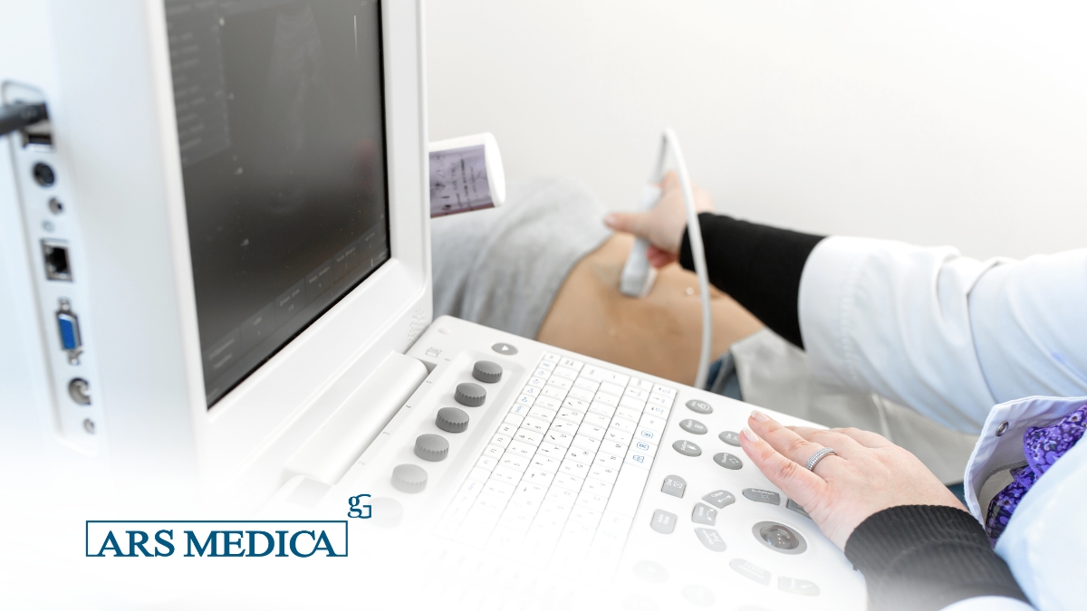
During the procedure, the specialist applies a water-based gel to the skin and glides a small probe (known as a transducer) over the area being evaluated. The transducer sends sound waves into the tissue, which bounce back as echoes that vary based on the density of the internal organs. The ultrasound machine’s computer processes these echoes in real time, converting them into dynamic images displayed on the screen.
Why Are Ultrasounds So Important?
An ultrasound often serves as the initial diagnostic checkpoint—providing the fastest and most efficient response to a clinical query. Above all, it is one of the most critical tools for preventive medicine and early cancer detection. Identifying a lesion in its earliest stages, often before noticeable symptoms appear, can dramatically improve treatment outcomes and recovery prognosis.
Key advantages include:
-
Early Detection: Enables the early identification of nodules, cysts, inflammatory processes, and structural anomalies within internal organs.
-
Real-Time Evaluation: Unlike static imaging, ultrasounds display organs in active motion (for instance, visualizing cardiac contractions or vascular blood flow via Echo-Color Doppler).
-
Zero Radiation Risk: Because there are no radiation-induced side effects, the scan can be repeated as often as necessary, making it ideal for tracking known conditions over time.
-
Procedural Guidance: Physicians frequently rely on real-time ultrasound imaging to guide joint injections and biopsies with sub-millimeter precision.
What Are the Primary Types of Ultrasound?
While ultrasound technology was once limited to evaluating solid abdominal organs and soft tissues, recent technological breakthroughs have expanded its capabilities. Today, clinicians routinely perform ultrasounds to evaluate specific pulmonary and intestinal conditions—areas previously considered inaccessible to acoustic imaging.
Common ultrasound applications include:
-
Abdominal Ultrasound (Complete, Upper, or Lower): Used to evaluate the liver, gallbladder, pancreas, spleen, kidneys, bladder, uterus, and prostate.
-
Bowel/Intestinal Ultrasound: An increasingly valuable tool for monitoring chronic inflammatory bowel diseases (such as Crohn’s disease and ulcerative colitis) and assessing intestinal wall thickness.
-
Thyroid and Neck Ultrasound: Essential for screening thyroid nodules and evaluating cervical lymph nodes.
-
Breast Ultrasound: A vital component of senology screening and early breast cancer detection, particularly in younger women, often used alongside mammography.
-
Lung Ultrasound: Highly effective for rapidly diagnosing pneumonia, pleural effusion, or interstitial lung disease without exposing the patient to radiation.
-
Musculoskeletal Ultrasound: Evaluates muscles, tendons, and joints following acute trauma or in cases of chronic pain.
-
Echo-Color Doppler: A specialized vascular ultrasound that assesses blood flow through arteries and veins, serving as a critical tool in preventing strokes, deep vein thrombosis, and venous insufficiency.
What Do You Need to Know Before Your Scan?
Many types of ultrasounds—such as thyroid, breast, lung, and musculoskeletal scans—require no special preparation. However, for abdominal and pelvic examinations, following pre-test instructions is essential for diagnostic accuracy:
-
Upper Abdominal / Intestinal Ultrasound: Requires fasting for at least 6 to 8 hours prior to the test. This prevents digestive activity and intestinal gas from obscuring underlying organs (particularly the gallbladder and pancreas).
-
Lower Abdominal / Pelvic Ultrasound: Requires a full bladder. Patients are generally advised to drink approximately one liter of plain water one hour before the appointment and refrain from urinating. A full bladder acts as an “acoustic window,” allowing clear visualization of the uterus, ovaries, or prostate.
The Role of the Specialist: Technology Is Only Half the Equation
There is a vital detail every patient should keep in mind: ultrasound imaging is highly operator-dependent. This means the accuracy of a diagnosis relies not only on state-of-the-art equipment, but heavily on the clinical expertise, diagnostic acumen, and skill of the specialist operating the transducer.
At our clinic, we combine cutting-edge imaging technology with a highly qualified team of ultrasound specialists, ensuring precise diagnostic results in a prompt, comfortable setting.